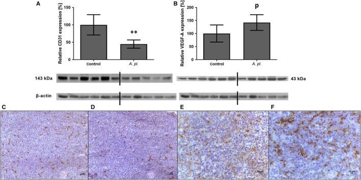
Figure 2.

Protein expression of CD31 and VEGF‐A in human pancreatic tumours grown in athymic mice treated orally with Arthrospira platensis for 2 wk. Western blot analysis of CD31 (A) and VEGF‐A expression (B), and representative microphotographs of CD31 immunohistochemical staining of control (C) and A platensis‐treated mice (D) and VEGF‐A staining of control (E) and A platensis‐treated mice (F). Densitometric quantification of immunoreactive bands was performed by recalculation to the β‐actin signal. The positivity for both antibodies is visualized by brown staining and counterstaining by haematoxylin. Bar 1 μm and 10 μm. Data in (A) and (B) represent median ± IQ range, p P = .056 vs control; **P < .01 vs control. The Western blot lanes represent the individual mouse tumour lysates. VEGF‐A, vascular endothelial growth factor A; A pl., Arthrospira platensis
