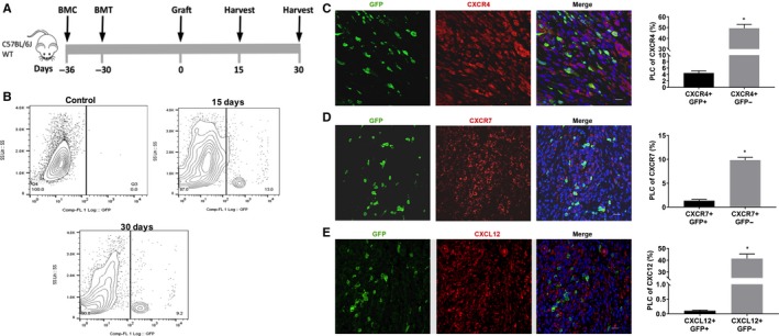
Figure 1.

A model of 5‐FU submyeloablation to characterize the bone marrow cell trafficking into endometriotic lesions in mice. Schematic model: Treatment for bone marrow conditioning (BMC) started on day −36 before uterine fragment grafting and 6 days before bone marrow transplantation (BMT) (A). Ectopic lesions were harvested 15 and 30 days (each group n = 6) after grafting uterine fragments into the peritoneal. Representative images of flow cytometry analysis of ectopic lesion cells on day 15 and 30 after uterine fragment grafting demonstrating the percentage of GFP‐positive cells in endometriosis. B, Control group (n = 6 mice), represented by GFP‐negative endometriosis lesions, was used to set a gate. Fluorescence confocal microscopy analysis of mouse endometriosis sections (C, D and E). Representative images of IF and quantitative analyses of cells expressing CXCR4, CXCR7 or CXCL12 and GFP, as a per cent of total endometriosis cells. Murine lesions were stained with anti‐GFP antibody (green) and co‐stained with CXCR4 antibody (red) (C), with CXCR7 antibody (red) (D) and with CXCL12 antibody (red) (E). Nuclei were stained by DAPI and are shown in blue. BMC, Bone morrow conditioning; BMT, Bone morrow transplantation; PLC, percentage of labelled cells. Results shown as mean ± SE. *P < .05 vs vehicle. Scale bar: 20 μm
