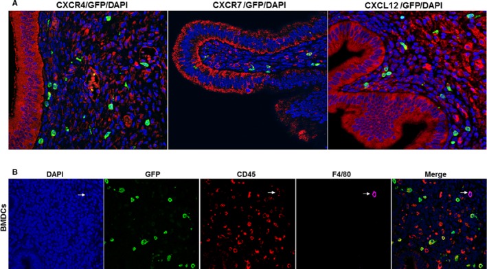
Figure 2.

Immunostaining of CXCR4, CXCR7 and CXCL12 and BMDCs. A, Representative immunofluorescence images of CXCR4, CXCR7 and CXCL12 staining in the epithelial cells of murine endometriosis. In our model, no GFP (bone marrow‐derived cells) was identified in the epithelium. Scale bar: 10 μm. B, Engrafted BMDCs express CD45 and F4/80. BMDCs: Engrafted bone marrow‐derived immune cells (BMDCs) were stained with DAPI or immunofluorescence for CD45 and F4/80. Engrafted BMDCs shows the presence of non‐immune (CD45 negative) and immune cells (CD45 positive), some of which co‐express F4/80, a marker of macrophages. Included is a cell that is a bone marrow‐derived (GFP+), CD45+, F4/80+ macrophage (arrow) Scale bar: 10 μm
