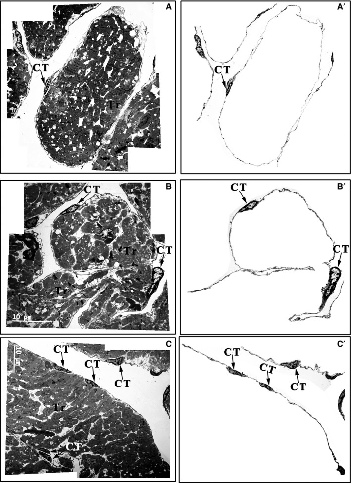
Figure 3.

Distribution of CTs in the upper region, middle region and base of the Xenopus tropicalis myocardium. CTs mainly concentrated on the outer surface of trabeculae containing cardiomyocytes in the upper region (A), middle region (B) and base (C) of the X tropicalis myocardium. Most of the trabeculae are twined around one, two or several CTs and their telopodes or with telopodes alone. A′, B′ and C′: CTs of A, B and C that are not included in the trabecular structure; CT: Cardiac telocyte; Scale bar: Size as shown in the figures; Tr: Trabecula of the myocardium
