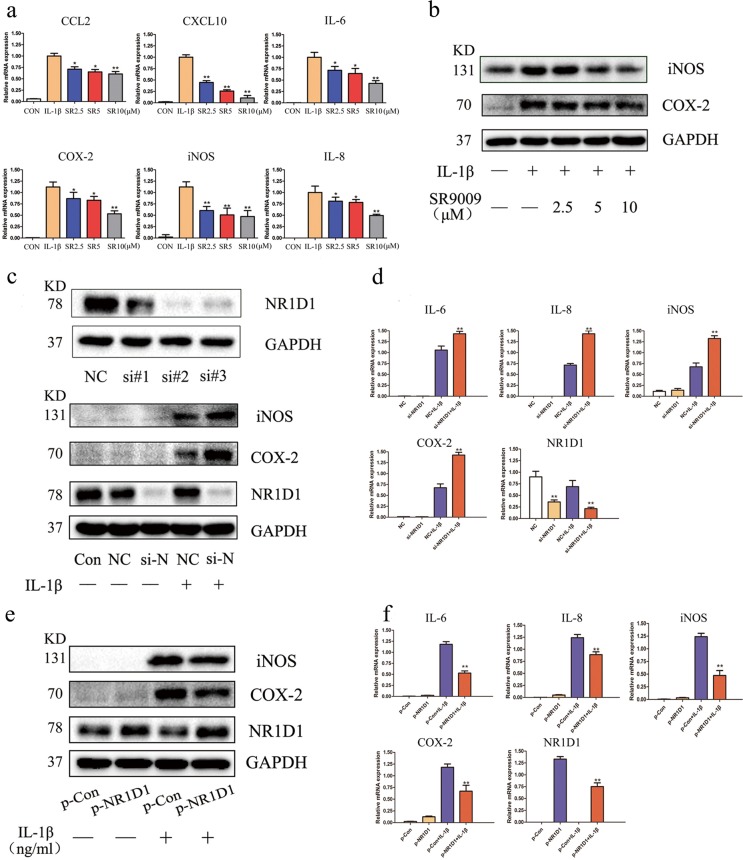
Fig. 2. NR1D1 regulates the inflammatory response in RA FLSs.
a IL-1β-mediated production of CCL2, CXCL10, IL-6, COX-2, iNOS, and IL-8 was diminished by SR9009, as determined by RT-PCR. Data are means ± SEM of three independent experiments. *p < 0.05, **p < 0.01 versus cells stimulated with only IL-1β. b COX-2 and iNOS levels were elevated by IL-1β but suppressed by SR9009, as determined by western blotting. c Efficacy of siRNA transfection. Total protein was extracted 72 h after transfection of RA FLSs with siRNA, and western blotting was performed (top). RA FLSs transfected with NR1D1 siRNA#2 exhibited increased COX-2 and iNOS levels after incubation with IL-1β (10 ng/mL). RA FLSs were transfected with NR1D1 siRNA and stimulated with IL-1β (10 ng/mL) for 72 h (bottom). d Expression of IL-6, IL-8, COX-2, and iNOS was increased in RA FLSs transfected with NR1D1 siRNA after incubation with IL-1β (10 ng/mL), as determined by RT-PCR. Data are means ± SEM of three independent experiments. *p < 0.05, **p < 0.01 versus negative control (NC) IL-1β-stimulated cells. e RA FLSs transfected with an NR1D1 overexpression plasmid exhibited decreased COX-2 and iNOS levels after incubation with IL-1β (10 ng/mL) as determined by western blotting. f Expression of IL-6, IL-8, COX-2, and iNOS decreased in RA FLSs transfected with an NR1D1 overexpression plasmid after incubation with IL-1β (10 ng/mL) as determined by RT-PCR. Data are means ± SEM of three independent experiments. *p < 0.05, **p < 0.01 versus empty plasmid and IL-1β-stimulated cells. si-N, si-NR1D1.

