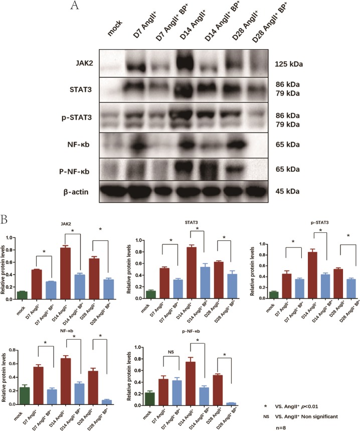
Fig. 4. BP-1-102 reduced the expression of inflammation signaling pathways-related proteins in AAA tissues.
a Western blotting was applied to analyze inflammation signaling pathways-related proteins expressions in AAA tissue at indicated time points. JAK2, STAT3, p-STAT3, NF-κB, and p-NF-κB expressions were evaluated in AngII+ group and compared with AngII+ BP-1-102+ group (AngII+BP+). AngII+ BP-1-102+ group showed reduced inflammation signaling pathways-related proteins compared with AngII+ group at each time point. Saline treated group was used as control (mock). b Relative protein levels of JAK2, STAT3, p-STAT3, NF-κB, p-NF-κB were determined by normalized with β-actin expression in AngII+ group and AngII+ BP-1-102+ group (AngII+BP+) at indicated time points. Saline treated group was used as control (green column). AngII+ BP-1-102+ group (blue column) showed reduced inflammation signaling pathways-related proteins compared with AngII+ group (red column) at each time point. The results were presented as mean ± standard deviation (SD). n = 8. *P < 0.01 vs. AngII+ group (two-way ANOVA followed by Tukey’s test). NS indicates non-significant differences compared with AngII+ group.

