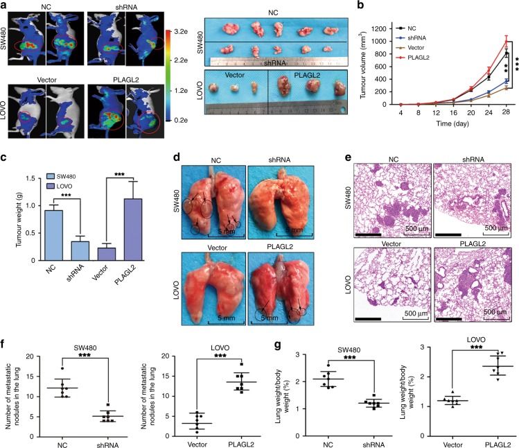
Fig. 3. PLAGL2 promotes CRC cell proliferation and metastasis in vivo.
a Representative bioluminescence pictures of the nude mice 28 days after injection. In all, 5 × 10⁶ stable SW480 and LOVO cells were injected subcutaneously into the groin of nude mice (n = 7 per group). Representative images of the corresponding xenograft 28 days after inoculation. b Tumour volumes in the different groups. The growth curve of the tumours that formed after subcutaneous injection. c Enhanced PLAGL2 expression increased the tumour weights. d Lung metastasis models. Representative images of visible lung metastases. The metastatic nodules are indicated with arrows. Scale bars, 5 mm. e Representative images of the corresponding HE staining. Scale bars, 500 μm. f The numbers of metastatic nodules. g The depletion of PLAGL2 significantly reduced the lung weight. *P < 0.05, **P < 0.01, ***P < 0.001, based on Student’s t-test.

