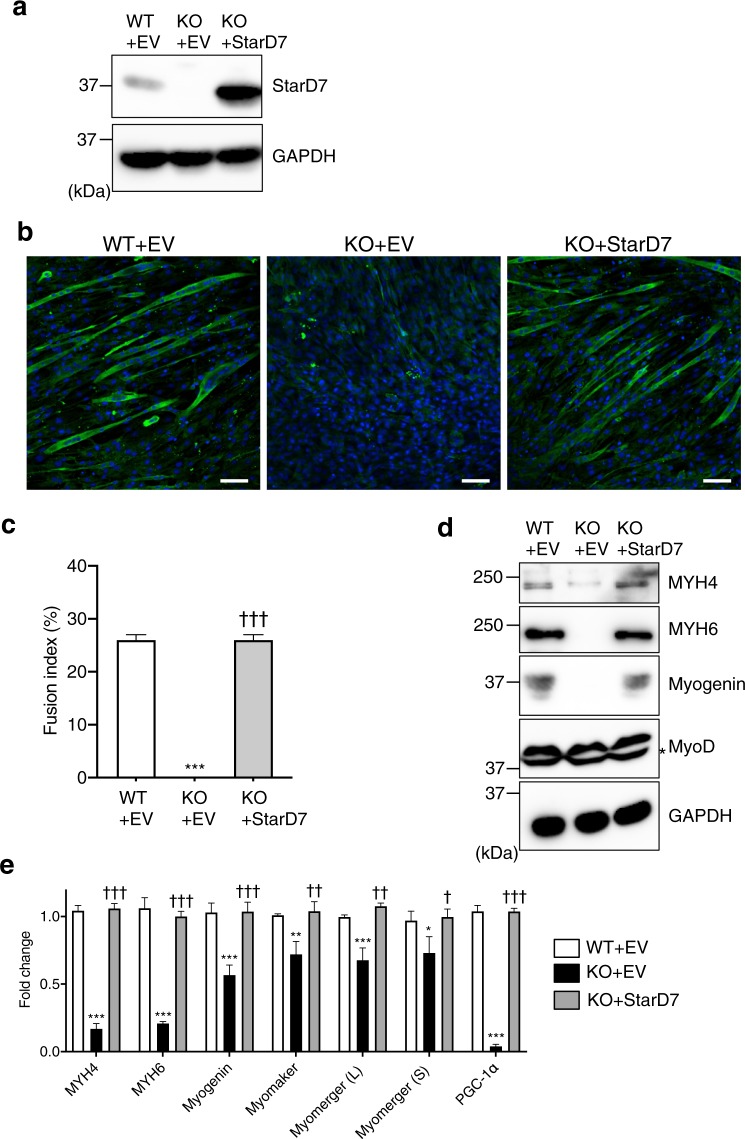
Figure 4.
Reintroduction of StarD7 into the StarD7-KO C2C12 cells restored myogenic differentiation. (a) Cell lysates from WT + EV (empty vector), KO + EV, and KO + StarD7 C2C12 cells were separated by SDS-PAGE and analyzed by western blotting using anti-StarD7 antibody. GAPDH was used as a protein loading control. (b) WT + EV, KO + EV, and KO + StarD7 C2C12 cells were cultured in differentiation medium for 5 days, then immunostained with anti-MYH6 antibody (green). Nuclei were stained with DAPI (blue). Bars indicate 50 μm. (c) The fusion indexes of C2C12 cells were calculated, and are presented as the means ± S.D. ***P < 0.001 as compared with WT + EV cells, and †††P < 0.001 as compared with KO + EV cells (one-way ANOVA with Tukey’s post hoc test). (d) After inducing differentiation, the amounts of MYH4, MYH6, myogenin and myoD protein were analyzed by western blotting. Asterisk indicates a non-specific band. (e) The mRNA levels of MYH4, MYH6, myogenin, myomaker, myomerger (L), myomerger (S) and PGC-1α were quantified by qPCR after inducing differentiation. Data were normalized to the GAPDH level. Values shown are means ± S.D. from three independent culture dishes. *P < 0.05, **P < 0.01, and ***P < 0.001 as compared with WT + EV cells, and †P < 0.05, ††P < 0.01, and †††P < 0.001 as compared with KO + EV cells (one-way ANOVA with Tukey’s post hoc test).

