
Figure 3.
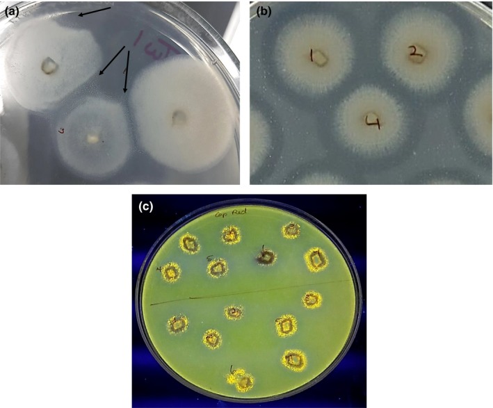
Lipid substrate bioassays; (a) arrows point to white crystalline precipitate on Tween‐20 medium; (b) zone of clearance (halo) around colonies producing secreted lipase (alpha/beta hydrolase/cutinase) on tributyrin medium; (c) yellow–orange fluorescent colonies on Rhodamine medium