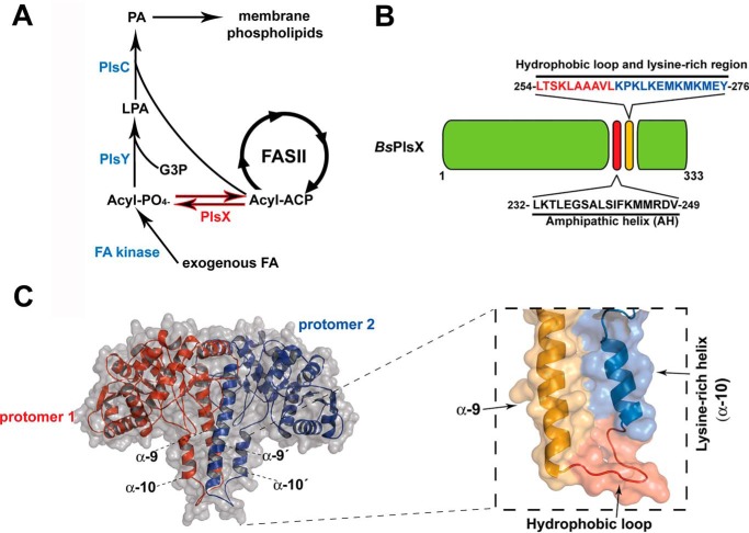
Figure 1.
Phospholipid synthesis in bacteria and the structure of PlsX. A, phospholipid synthesis pathway in Gram-positive bacteria. B, diagram of the B. subtilis PlsX protein showing the position of the putative AH (232–249) and the hydrophobic exposed region (254–263) and lysine-rich region (264–276). C, right, structural model of B. subtilis PlsX dimer performed by Swiss Model homology-modeling server (31) (using PDB 1VI1 as a template), indicating the localization of the hydrophobic loop and the lysine-rich helix α10. Left, zoomed-in view of the α9 and α10 helices in a PlsX monomer.

