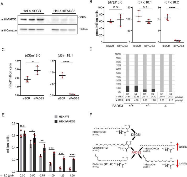
Figure 3.
FADS3 is required for Δ14Z LCB desaturation. A, HeLa cells transfected with siFADS3 or siSCR. The silencing of hFADS3 was confirmed by Western blotting with a polyclonal antibody against FADS3. Calnexin was used as a loading control (n = 3). B–D, sphingoid base profiling of cells and plasma. B, HeLa cells were transfected with siSCR and siFADS3 for 72 h and subsequently cultured for 24 h in the presence of 2 μm (d7)d18:0 and (d3)m18:0. The formation of (d7)d18:2 was significantly decreased in the siFADS3-treated cells. No difference was seen for the precursors (d7)d18:0 and (d7)d18:1. C, the conversion of (d3)m18:0 into (d3)m18:1 was also reduced in siFADS3-treated cells compared with controls (siSCR). Data are shown as mean ± S.D. (error bars), unpaired t test; *, p < 0.05; ***, p < 0.001. D, d18:2 was absent in plasma of the two FADS(−/−) mice (M1/M2), and about 50% in the FADS(+/−) mice compared with WT. Total d18:1 levels were not different. E, WT and FADS3-overexpressing HEK293 cells were plated at low density (150,000 cells/well of a 24-well plate) and supplemented with increasing concentrations of m18:0 (0–1.5 μm). After 48 h, the number of attached cells was significantly higher in FADS3-overexpressing cells than in WT. Data are shown as mean ± S.D. (n = 3) and compared by two-way analysis of variance with Bonferroni's correction *, p < 0.05; **, p < 0.01; ***, p < 0.001; n.s., not significant. F, interplay between the two LCB desaturases DEGS1 and FADS3. DEGS1 introduces the Δ4E DB into d18:0, forming d18:1, whereas FADS3 introduces the Δ14Z DB into d18:1 and m18:0.

