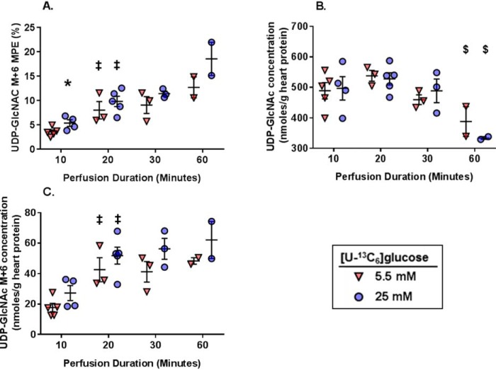
Figure 8.
The effect of [U-13C6]glucose concentrations and perfusion durations on 13C-labeling and concentration of myocardial tissue metabolites relevant to the HBP. A, UDP-GlcNAc M+6 MPE. B, UDP-GlcNAc concentrations. C, UDP-GlcNAc M+6 myocardial concentrations. For all graphs, n = 5 for 5.5 mm 10-min perfusions, n = 4 for 25 mm 10-min perfusions, n = 3 for 5.5 mm 20-min perfusions, n = 5 for 25 mm 20-min perfusions, n = 3 for 5.5 mm 30-min perfusions, n = 3 for 25 mm 30-min perfusions, n = 2 for the 5.5 mm 60-min perfusions, n = 2 for the 25 mm 60-min perfusions. *, p < 0.05 between the groups at the same perfusion duration; ‡, p < 0.05 versus the immediately preceding perfusion duration for the same glucose concentration; $, p < 0.05 versus the 20-min perfusion duration for the same glucose concentration.

