
Figure 6.
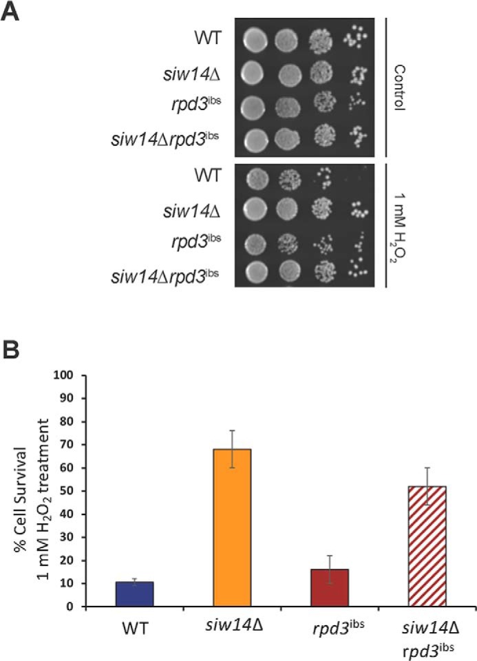
The siw14Δ mutation is epistatic to the rpd3ibs mutation. A, WT, siw14Δ, rpd3ibs, and siw14Δ rpd3ibs strains were grown to mid-log phase and treated with 1 mm H2O2 for 3 h, as described in the legend to Fig. 1. Cells were plated and incubated at 30 °C for 2 days. Colony-forming units were determined, and the percent survival was calculated (cfu treated/cfu untreated, ×100). B, bars represent the average of 9 biological replicates over 3 assays. Error bars represent mean ± S.D. **, p value ≤ 0.001.