Figure 2.

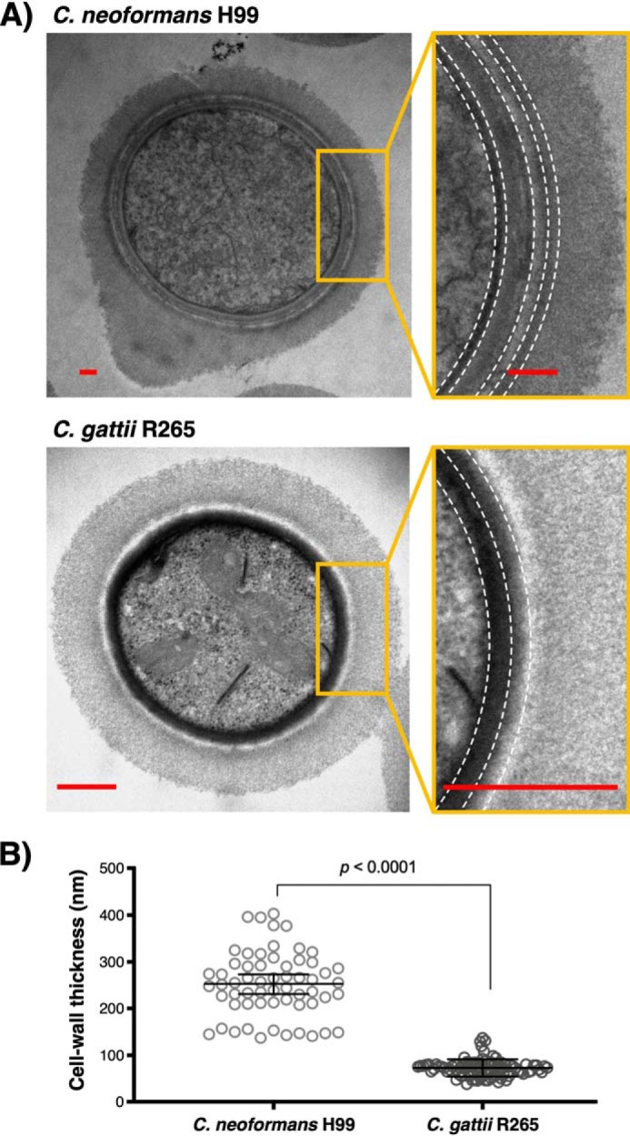
The melanized cell wall of C. gattii R265 reveals a compact and uniform deposition of melanin pigments. A, representative cross-sectional transmission electron micrographs of C. neoformans H99 (top) and C. gattii R265 (bottom) melanized yeast cells. The magnified images show a close-up view of the cell wall with dashed lines to illustrate the difference in layered melanin pigment arrangement between these two cryptococcal isolates, possibly attributable to their cell-wall chitinous compositions. Scale bar, 500 nm. B, measurement of C. neoformans H99 and C. gattii R265 melanized cell-wall thickness. Data were obtained by measuring 12–18 cells per strain (5 measurements/cell) and analyzed using the Mann–Whitney test, two-tailed p value with 99% confidence level. Error bars, 95% confidence interval of the median.
