Figure 8.
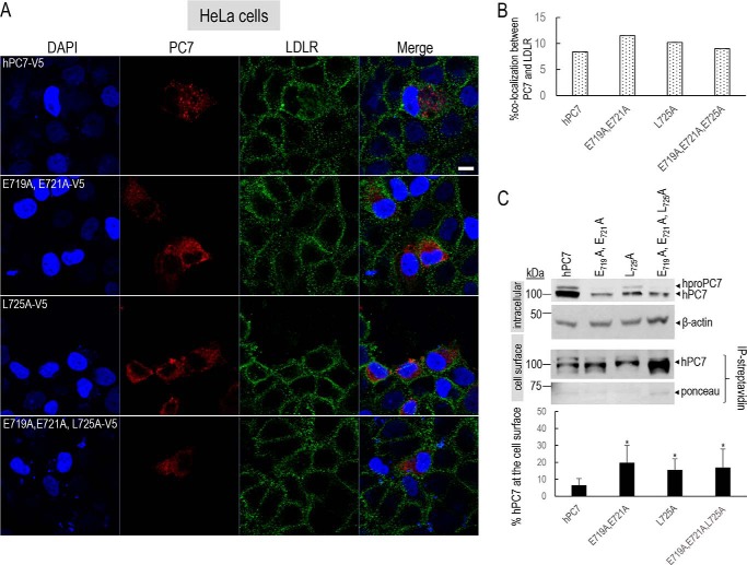
Localization of human PC7 and its CT mutants at the cell surface. A, immunofluorescence of hPC7 or its CT mutants (red) and LDLR (green) in nonpermeabilized HeLa cells. Cell nuclei are marked by DAPI (blue). B, quantification of the immunoreactivity of hPC7 or its mutants with the cell-surface marker LDLR using IMARIS software. These results are representative of a minimum three independent experiments, and quantification represents an average of n = 15 cells per condition. C, Western blot analysis of PC7 WT and mutants, biotinylated and immunoprecipitated with streptavidin beads. Error bars indicate averaged values ± S.D. *, p < 0.1, (Student's t test). Bar = 1 μm.

