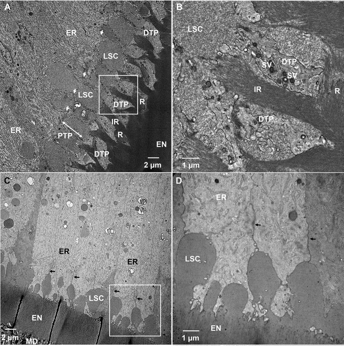
Figure 10.
TEM micrographs of demineralized and resin-embedded sections of WT and KI incisors during the secretory stage of enamel formation. A, WT secretory stage ameloblasts. B, close up of the boxed area in A. C, KI early secretory stage ameloblasts. D, close up of boxed area in C. EN, enamel; ER, rough endoplasmic reticulum; DTP, distal Tomes' process; IR, interrod; LSC, large secretory compartments; MD, mantle dentin; PTP, proximal Tomes' process; arrows, distal junctional complexes; double-headed arrow, area of PTP in WT secretory ameloblasts; SV, secretory vesicles.

