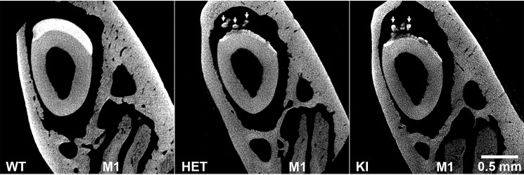
Figure 6.
μCT analyses of mature enamel from WT, HET, and KI enamel. Analyses show that KI enamel layers at maturation are under-mineralized compared with the thick WT enamel layer. The example of the HET enamel shown is similar to KI enamel. As discussed, KI enamel and some HET enamel specimens (as shown) exhibit ectopic mineral deposits (arrows). Note the presence of molar M1 roots, indicative of the enamel maturation stage. These images were obtained using an Xradia MicroXCT-200 instrument (see “Experimental procedures”).

