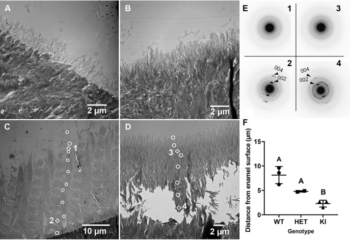
Figure 7.
Selected TEM images and SAED results of the developing enamel matrix of WT and KI maxillary incisors. A, early secretory stage (grid 1) in WT enamel exhibiting a rodless enamel layer on more densely mineralized dentin. B, early secretory stage of enamel formation (grid 1) in KI enamel, where a rodless enamel layer is again observed, as seen in WT enamel (A). C, mid-secretory stage (grid 5) in WT enamel. A thicker enamel layer with an enamel rod structure is clearly observed. White circle and diamond-shaped markers represent locations of SAED measurements. SAED results at diamond-shaped marker locations are presented in E. Additional results are found in Fig. S4. D, mid-secretory stage (grid 4) of KI enamel, as throughout KI enamel development, grows perpendicular to the DEJ and lacks an enamel rod structure. E, selected SAED findings near the enamel surface (C, location 1; D, location 3) and close to the DEJ (C, location 2; D, location 4). F, comparison of rates of ACP transformation to apatitic crystals in developing WT (n = 3), HET (n = 2), and KI (n = 3) enamel. Mean ± S.D. with different letter notations are significantly different (p < 0.05). The biological and mechanistic importance of noted differences in ACP stabilization and rates of mineral phase transformation are discussed in the text.

