
Figure 2.
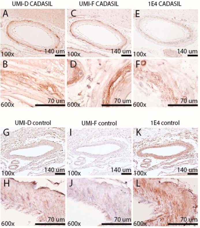
Comparison of NOTCH3 staining by monoclonal antibodies UMI-D and UMI-F and a standard NOTCH3 antibody (1E4, EMD Millipore). UMI-D (A, B, G, and H), UMI-F (C, D, I, and J), and 1E4 (E, F, K, and L) were used for immunohistochemical localization of NOTCH3 antigen in the frontal lobe of four genetically verified CADASIL (A–F) and four control brains (G–L). Representative photographs were captured from serial sections of representative leptomeningeal vessels at the magnifications shown. UMI-D and UMI-F (A–D) reacted more strongly with NOTCH3 in CADASIL leptomeningeal vessels, compared with 1E4 (E and F). UMI-D and UMI-F demonstrated little to no staining in the medial layers of cerebral arteries in control patients (G–J), whereas 1E4 demonstrated increased staining of control vessels (K and L).