Figure 7.
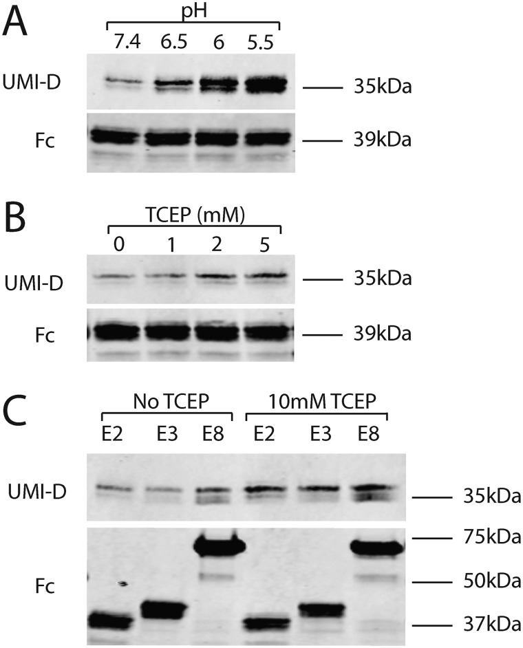
The effect of pH and redox state on fragmentation of NOTCH3 at Asp80. A, purified protein (Fc-E2) was exposed to buffers at specific pH for 1 h at 37 ºC, boiled in reducing sample buffer, and electrophoresed on gels. Western blot analysis was performed using UMI-D to detect fragmentation (top) and Fc to detect total protein input (bottom). There was a clear increase in fragmentation at modestly acidic conditions. B, the same purified protein was exposed to a series of TCEP concentrations for 1 h at 37 ºC and analyzed as in A. There was a consistent increase in fragmentation with increasing concentration of TCEP. C, purified proteins with different numbers of EGF-like repeats were exposed to TCEP or vehicle prior to Western blot analysis as in A. All proteins exhibited increased fragmentation with TCEP incubation. All experiments were performed four or more times and showed the same findings.

