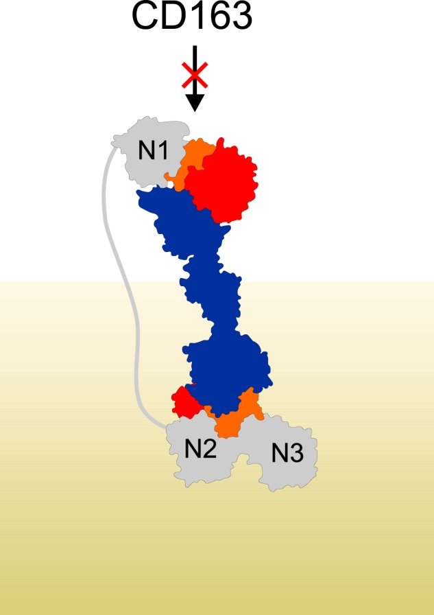
Figure 7.

IsdHN1 blocks interaction between Hp–Hb and CD163. Shown is a model of IsdHN1-N3 binding to Hp–Hb. The model was generated based on the structures of HpSP–αβHb–IsdHN2-N3 and Hp–Hb–IsdHN1 (PDB entry 4WJG) (36). The distance between IsdHN1 C terminus and IsdHN2 N terminus is ∼180 Å. IsdH is shown in gray, Hp is in blue, αHb is in orange, and βHb is in red. The peptidoglycan layer on the surface of the bacterium is indicated in khaki.
