Figure 3.
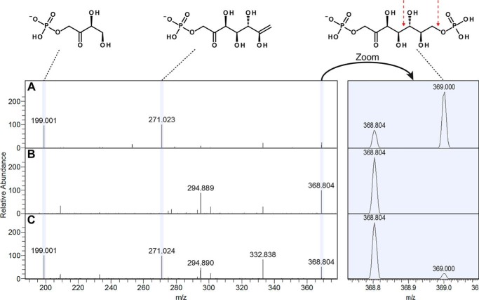
Identification of SBP (m/z 368.99) via targeted monitoring of fragments for the precursor ion at m/z 368–370. A, E. coli ΔtalAB extract; B, blank run; C, xylose grown C. thermosuccinogenes extract. A clear peak is present in A and C at m/z 369 corresponding to SBP, confirmed by indicative fragment peaks at m/z 199 and 271, which are further absent in the blank (B). The red arrow indicates the bonds that, when broken, result in the fragment at m/z 199 and 271. The identity of the peak at m/z 368.804 is unknown. Presence of this peak in the blank run (B) indicates that it is a background signal, whereas the nearby peak at m/z 369 is not.

