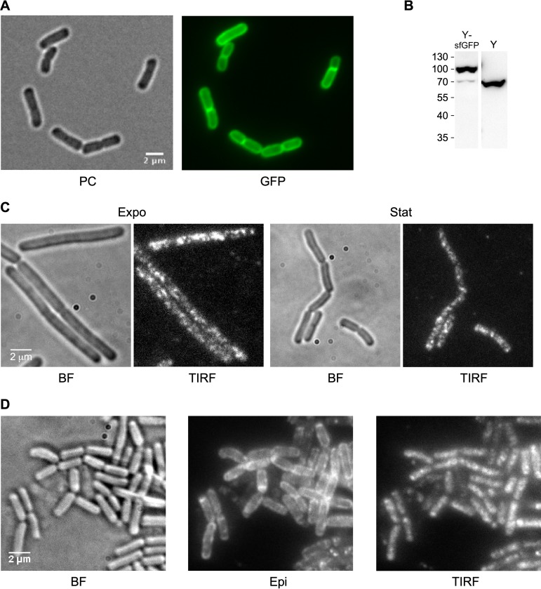
FIG 1.
Visualization of RNase Y in the cell. (A) RNase Y localizes at the membrane. Strain SSB2063a (Pxyl-rny-mgfpmut1) was grown in SMS minimal medium at 37°C to early stationary phase in the presence of 60 mM xylose to induce expression of the RNase Y-GFP fusion protein and observed by phase-contrast (PC) and wide-field fluorescence microscopy (GFP). (B) Quantification of RNase Y-sfGFP and RNase Y expression by Western blotting. Total cell extracts of strains SSB2048 and SSB1002 (WT) were analyzed using an anti-RNase Y monoclonal antibody. For further details, see the legend to Fig. 6D. (C) RNase Y focus formation at the membrane. Cells of strain SSB2063a grown in LB medium at 37°C and expressing RNase Y-GFP fusion protein were imaged at mid-log (Expo) and stationary phase (Stat). The focus pattern of RNase Y-GFP fusion protein was visualized by TIRF microscopy (100-ms exposure time). BF, bright field. (D) RNase Y-sfGFP (construct at the rny locus, SSB2048) dynamically localizes in membrane foci. The cultures were grown to mid-log phase in the SMS minimal medium at 37°C. The TIRF images were extracted from 30-s time-lapse movies. All images were acquired on living cells deposited on an agar pad and covered by a glass slide. Scale bar, 2 μm. BF, bright field; Epi, epifluorescence; TIRF, total internal reflection fluorescence.

