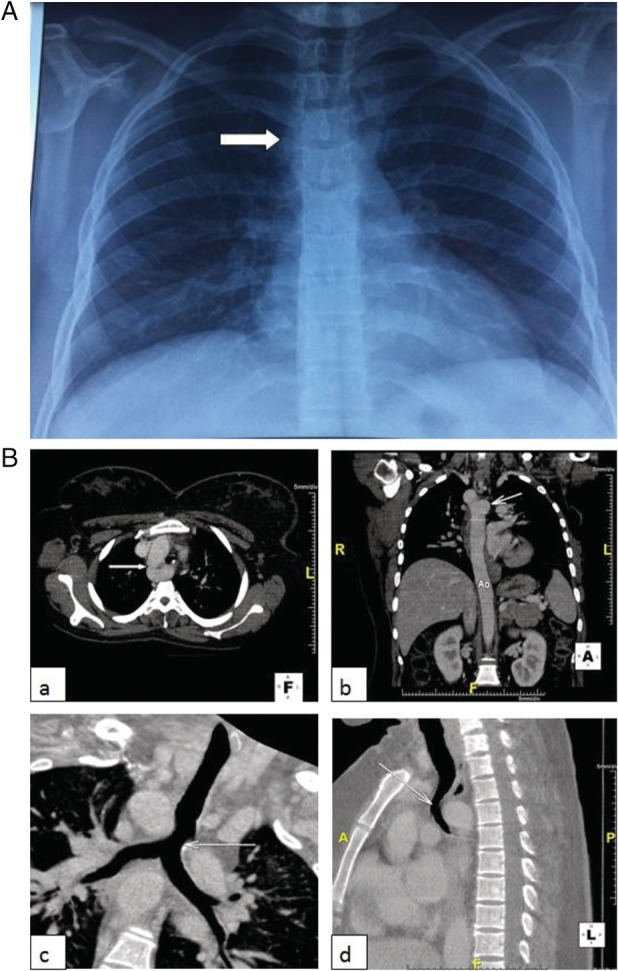
Figure 1.

Imaging findings: (A) Frontal chest radiograph: right‐sided aortic arch with a right‐sided aortic knob (white arrow). (B) Chest CT Scan: a right‐sided aortic arch and a tracheal compression due to the diverticulum of Kommerell. (a) Axial image: right‐sided aortic arch (white arrow). (b) Coronal image: diverticulum of Kommerell (white arrow). (c) Coronal image (reconstruction): tracheal compression (white arrow). (d) Sagittal image: tracheal compression (white arrow).
