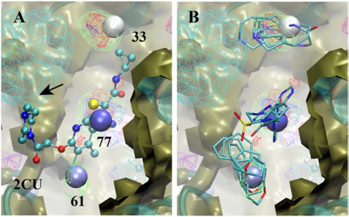
Figure 10).
Allosteric binding site of the M2 muscarinic receptor showing the A) crystallographic orientation of allosteric modulator 2CU (CPK) and B) selected fragments from the Hotspots analysis. Included are the SILCS exclusion maps (tan, solid surface), protein backbone (cyan, cartoon representation), the three Hotspots (vdW spheres, coloring based on mean LGFE scores, Table 3), and selected SILCS FragMaps with cutoff energies for visualization: Positive (cyan, −1.2 kcal/mol), Negative (orange, −1.2 kcal/mol), Apolar (green, −1.2 kcal/mol), H-bond donor (blue, −0.9 kcal/mol) and H-bond acceptor (−0.9 kcal/mol). Fragments shown include 3, 37, 41, 45 and 84 for site 33, 6, 49, 61, 76 and 83 for site 66, and 4b, 52b, 63c and 88 for site 77 (Figure S1 supporting information).

