Fig. 1.

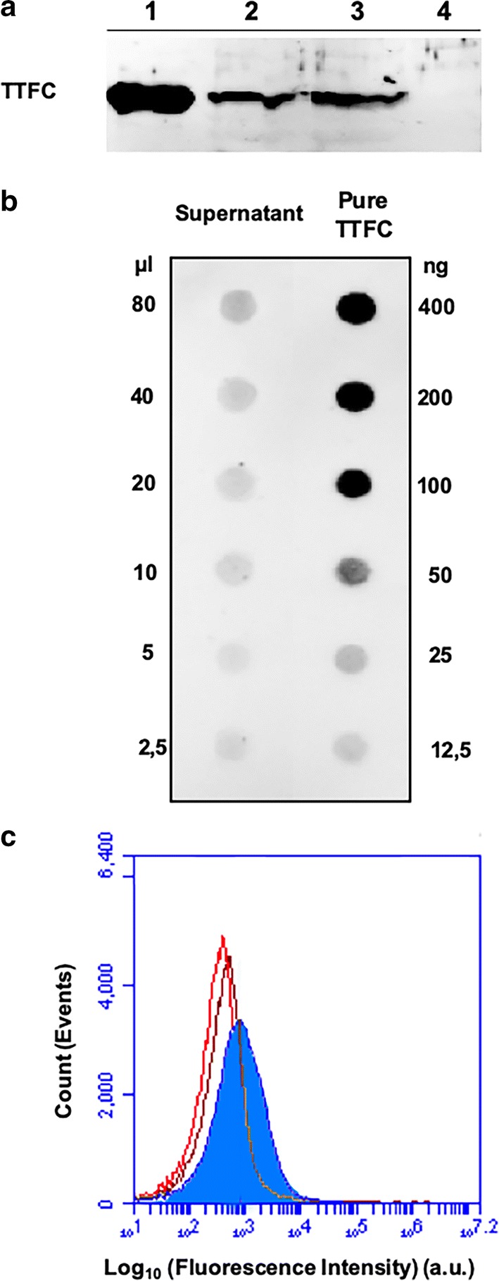
TTFC adsorption on B. subtilis spores. a Western blotting of spore surface proteins after adsorption with 2.0 µg of purified TTFC. Lanes 1: purified TTFC; 2: proteins extracted from adsorbed spores; 3: proteins extracted from adsorbed spores after 1 week storage at 4 °C; 4: five-fold concentrated supernatant after 1 week storage at 4 °C. b Dot blotting experiment performed with the serial dilutions of the supernatant (unbound TTFC) fraction of the adsorption reaction. Serial dilutions of purified TTFC were used as a standard. c Flow cytometry analysis of: free spores incubated (brown histogram) or not (red histogram) with specific antibodies and TTFC-adsorbed spores incubated with specific antibodies (filled blue histogram). The analysis was performed on the entire spore population (ungated). Immune-reactions were performed with polyclonal anti-TTFC [17] and anti-rabbit HRP conjugate (panels A and B) or with FITC-conjugated secondary antibodies (panel C)
