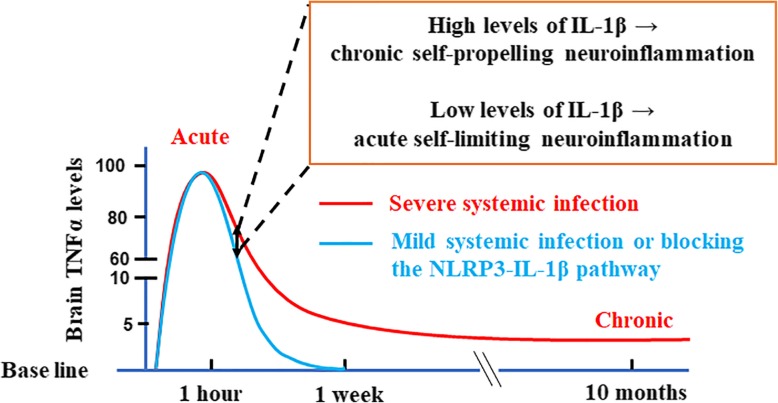
Fig. 8.
Schematic drawing depicting how brain IL-1β levels determine the fate of neuroinflammation induced by endotoxemia. Initial acute proinflammatory responses in the brain are similar between mild and severe endotoxemia; however, long-term outcomes are different. The curves of microglial activation are represented by TNFα concentrations in brain, because our previous study demonstrated that brain TNFα peaked at 1 h after LPS i.p. injection and then lasted at low levels in the endotoxemia-elicited neurodegeneration mouse model [17]. The NLRP3-IL-1β pathway gauges the severity of endotoxemia and generates dose-dependent increases of brain mature IL-1β around 7–11 h after LPS injection. Low levels of brain IL-1β permit the resolution of acute neuroinflammation as occurred in mild endotoxemia. In contrast, high levels of brain IL-1β during severe endotoxemia lead to chronic self-propelling neuroinflammation. This figure also suggests that in severe endotoxemia, reducing the production or hampering the function of brain IL-1β during the systemic acute inflammatory stage may provide a therapeutic window by preventing the transition of acute to chronic neuroinflammation and lowering the risk of resultant progressive neurodegeneration

