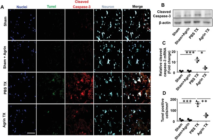
Figure 7.

Agrin treatment protecting neurons at 72 h after I/R injury. A) Cleaved caspase‐3 (red), Tunel (green), and Nissl staining were measured by confocal microscopy in the ipsilateral cortex (scale bar, 50 µm). B) Activated caspase‐3 expression was measured by western blotting and C) real‐time PCR in the ischemic penumbra (mean ± SD, n = 3, **p < 0.01). D) Quantitative analysis of the Tunel‐positive cells (mean ± SD, n = 3, **p < 0.01, ***p < 0.001).
