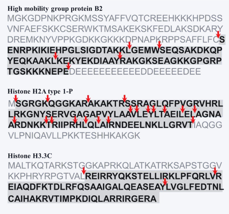
Figure 3.
Truncated proteoforms shown (highlighted in grey) against the sequences of three exemplary gene products. Red arrows indicate sites of truncation. Top: High mobility group protein B2. Six cleavage sites were identified in ten truncated proteoforms. Middle: Histone H2A type 1-P. Nineteen cleavage sites were identified in thirty-one truncated proteoforms. Bottom: Histone H3.3C. Three cleavage sites were identified in three truncated proteoforms.

