
Figure 3.
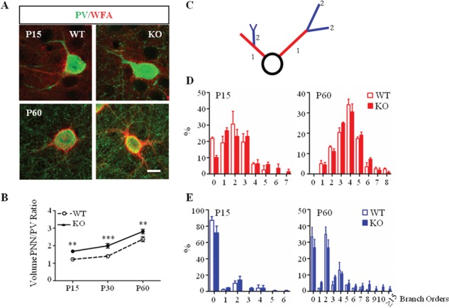
Increased complexity of PNNs enwrapping PV cells in Mecp2 KO mice. (A) Double immunostaining of PNNs with WFA (red) and PV cells (green) in immature (P15) and mature (P60) visual cortex of Mecp2 WT and KO mice. Scale bar: 10 μm. (B) Somata volume of PNN around PV-positive cell body increased in Mecp2 KO throughout animal life. (C) Schematic diagram showing classification of WFA-positive primary and secondary branches. (D) Percentage of primary PV-positive branches surrounded by WFA is increased in Mecp2 KO compared to littermates WT at P15. No differences at P60. (E) Percentage of secondary PV-positive branches is upregulated in Mecp2 KO both at P15 and P60. n = 5–6 animals per age and genotype. Two-way ANOVA, *P < 0.05; **P < 0.01; ***P < 0.001, Bonferroni’s post-test. Mean ± s.e.m.