
Figure 4.
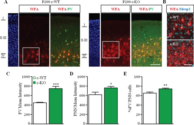
Mecp2 deletion in PV cells promotes the formation of PNNs around PV cells. (A) Triple staining for WFA (red), PV (green), and MeCP2 (cyan) in the visual cortex of Mecp2+/y/PV-Cre Het (c-WT) and Mecp2lox/y/PV-Cre Het (c-KO) at P200. (B) Selective deletion of Mecp2 in WFA-positive cells. (C, D) PV (C) and WFA (D) immunofluorescent mean intensity are upregulated in c-KO compared to c-WT littermates. (D) Percentage of PV-positive cells enwrapped by WFA is increased in c-KO compared to c-WT. Scale bar: 100 μm (A); 50 μm (B). n = 3 animals per genotype. Mann–Whitney test, *P < 0.05; **P < 0.01; ***P < 0.001. Mean ± s.e.m.