Figure 5.

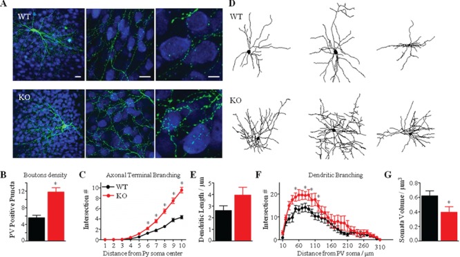
Axonal and dendritic hyper-ramification of PV cells in Mecp2 KO mice. (A) Mecp2 WT and KO cortical organotypic cultures were transfected via biolistics with a Pg67-GFP construct. A KO basket neuron (lower panel, green) among other NeuN immunostained neurons (blue) shows elaborate axonal branching and numerous boutons on the postsynaptic somata, as compared to a WT basket neuron (higher panel). Scale bar: 20 μm (left), 20 μm (middle), 5 μm (right). (B, C) Axonal bouton density (B) and complexity of terminal branching (C) are significantly increase in Mecp2 KO basket neurons (Mann–Whitney test, *P < 0.01). (D) Representative Neurolucida-traced basket cells in Mecp2 WT and KO animals. (E, F) Sholl-analysis reveals a significant increase in the total dendritic length (E) and in the complexity of dendritic branching (F) in Mecp2 KO. (G) PV-positive somata volume of Mecp2 KO basket cells is significantly reduced, as compared to WT littermates. n = 7 WT basket cells; n = 10 KO basket neurons. Mann–Whitney test, *P < 0.01. Mean ± s.e.m.
