Figure 7.

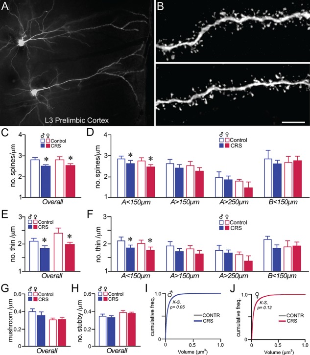
CRS induces dendritic spine loss in PL pyramidal neurons in male and female rats. (A) Darkfield photomicrograph illustrating examples of Lucifer Yellow dye-filled layer 3 pyramidal neurons in PL. (B) Examples of deconvolved images of dendritic segments from layer 2/3 pyramidal neurons in PL. Scale bar = 50 μm (A), 5 μm (B). (C, D) Mean + SEM for overall dendritic spine density (C) and density values within specific dendritic compartments (D). CRS decreased overall spine density in both sexes, and was accounted for by downward trends throughout all apical dendritic compartments, and significant decreases in the < 150-μm apical distance. (E, F) Both the overall density of thin spine subtypes (E) and density values within the specific dendritic compartments sampled (F) illustrate an attrition in this subtype following CRS that accounts for much of the overall decreases above. *, indicates significant difference between control and CVS; P < 0.05 (C, E), P < 0.0125 (D, F). N = 6–8/group. (G, H) CRS had no effect on mushroom (G) or stubby (H) subtypes in PL neurons. (I–J) Cumulative frequency distributions of overall spine volume in PL neurons do not reveal any significant population shifts in spine volume. K-S, Kolmogrov-Smirnov test, significance set at P < 0.01.
