
Figure 2.
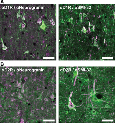
Immunofluorescent staining of dopamine receptors on neurogranin+ and SMI-32+ pyramidal neurons. (A) Left: many neurogranin+ pyramidal neurons (pink) express D1Rs (green). Right: the majority of SMI-32+ neurons (pink) also express D1Rs (green). (B) Left: several neurogranin+ pyramidal neurons (pink) express D2Rs (green). Right: the majority of SMI-32+ pyramidal neurons (pink) express D2Rs (green). Scale bar = 100 μm.