
Figure 5.
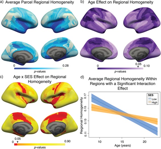
Variation in age and SES is associated with ReHo of functional brain networks at rest. (a) On average, values of ReHo are the largest in the inferior parietal lobe, precuneus, and posterior cingulate. (b) Regional P values for the effect of age on ReHo. Negative associations between age and ReHo were widespread and were strongest in the precuneus, inferior parietal lobe, and premotor cortex. (c) Regional P values for the effect of SES on associations between ReHo and age. Image is thresholded to control for multiple comparisons using the false discovery rate (q < 0.05); significant regions are shown in red. (d) SES effects on the associations between ReHo and age, extracted from only nodes that show a significant age-by-SES interaction.