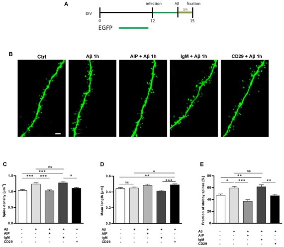
Figure 2.

Hippocampal slices treated with Aβ oligomers show an increase in the total dendritic spine density, which is reverted with CaMKII and integrin β1 inhibitors. (A) Organotypic hippocampal slices were infected with sindbis virus expressing EGFP at day 12 in vitro. Three days later, slices were treated with Aβ oligomers 1 μM or vehicle and fixed at day 15 in vitro as indicated on the timeline. (B) Micrographs obtained by confocal imaging show apical dendritic segments from CA1 hippocampal neurons in control and Aβ treatment conditions in the presence of different inhibitors, AIP (CaMKII inhibitor) and CD29 (integrin β1 inhibitor). Scale bar, 2 μm. (C,D) Bar graphs represent quantification of spine density and spine mean length in different cultures. (E) The bar graph shows quantification of stubby spine density after Aβ treatment in the presence or absence of inhibitors. *p < 0.05, **p < 0.01, ***p < 0.001, n.s. non-significant, compared to non-treated cells; paired one-way ANOVA. Data are represented as mean ± standard error of the mean (SEM), n = 35 dendrites of 5–6 neurons per condition.
