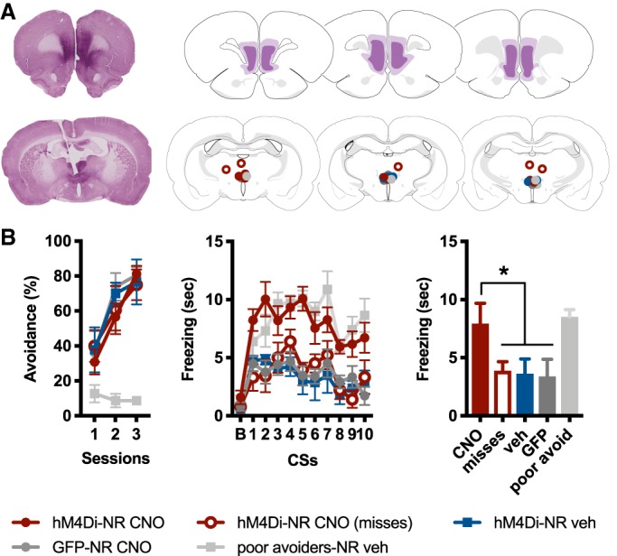
Figure 2.

(A) Representative example of hM4Di expression in vmPFC, visualized with VIP (top left). Greatest/least extent of hM4Di expression in vmPFC (top right). Representative example of cannula placement, with VIP-labeled vmPFC fibers evident in NR (bottom left). Cannula hits in and around NR (bottom right). Brain maps adapted from Swanson (2004). (B) Percent total possible avoidance responses across three daily sessions of SAA training (left); freezing during a 15-sec baseline (B), followed by the 10 CSs presented during test (middle); freezing averaged across the 10 test CSs (right). The hM4Di-NR CNO group showed enhanced freezing relative to other groups, with the exception of poor avoiders (*indicates P < 0.01).
