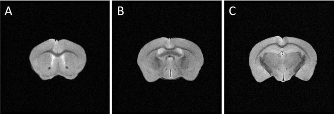
Figure 3.

T2-weighted axial magnetic resonance (MR) images of a mouse brain. Images acquired on a 14.1T microimager show slices containing the genu (A; bregma 0.98 mm), body (B; bregma −0.34 mm), and splenium (C; bregma −1.82 mm) of CC. Imaging parameters: TR/TE 2,000 ms/40 ms, 1 average, slice thickness 0.5 mm, field of view (FOV) 1.5 cm × 1.5 cm, matrix size 256 × 256. Ex vivo DTI was acquired from these slice locations with the same slice thickness and in-plane resolution (59 μm × 59 μm).
