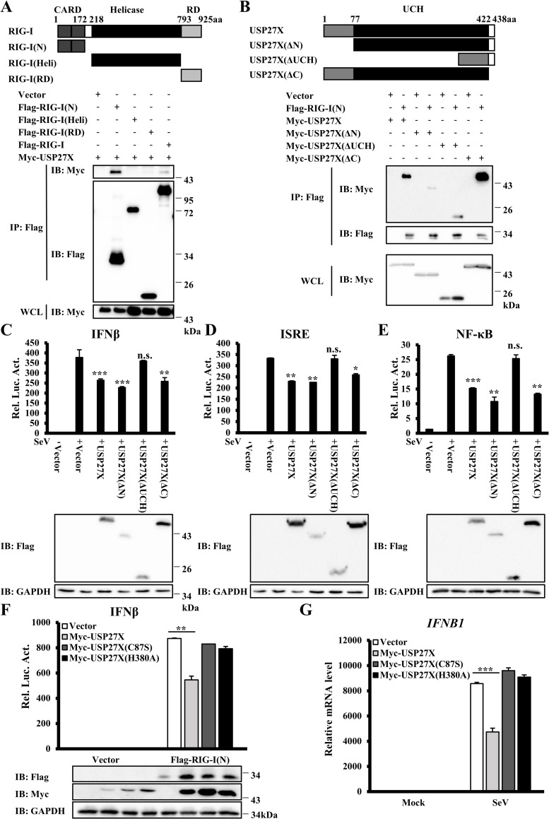
Fig 5. The USP27X UCH domain is essential for its inhibition of type I IFN signaling.
(A) HEK293T cells were transfected with the indicated expression plasmids. Cell lysates were immunoprecipitated with anti-Flag beads, followed by immunoblotting. (B) Similar to (A) except that HEK293T cells were transfected with different expression plasmids as indicated. (C–E) HEK293T cells were co-transfected with the indicated expression plasmids along with luciferase reporter constructs driven by promoters of IFNβ (C), ISRE (D) or NF-κB (E). Twenty-four hours after transfection, the cells were infected with SeV for 12 h. The cells were lysed for luciferase assays (upper panel) and immunoblotting assays (lower panels). (F) HEK293T cells were co-transfected with the indicated expression plasmids and IFNβ reporter. Twenty-four hours after transfection, the cells were lysed for luciferase assays (upper panel) and immunoblotting assays (lower panels). (G) HEK293T cells were transfected with the indicated expression plasmids. Twenty-four hours after transfection, the cells were infected with SeV for 9 h. The cells were lysed for measurement of IFNB1 mRNA levels by qRT-PCR. The data shown in C–G are from one representative experiment of at least three independent experiments [mean ± SD of duplicate experiments in (C–F) or triplicate experiments in (G)]. The two-tailed Student’s t-test was used to analyze statistical significance. * P < 0.05, **P < 0.01, ***P < 0.001, n.s. not significant versus control groups.

