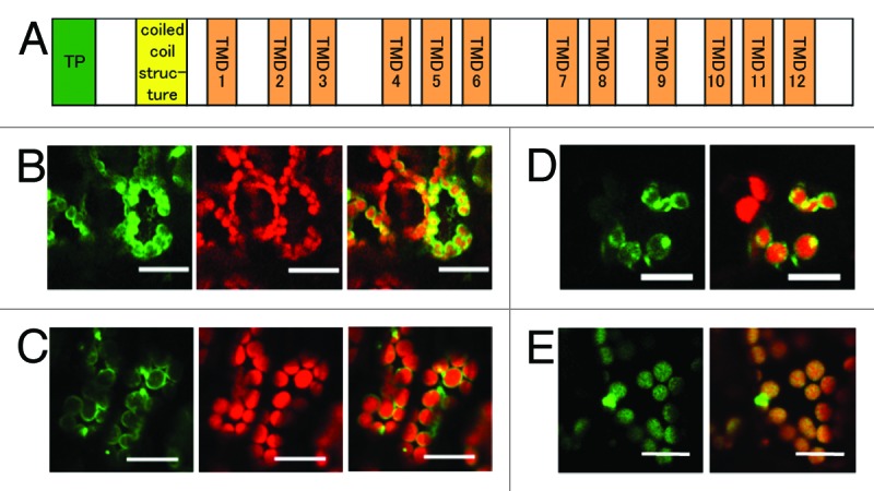
Figure 1. Localization of the EDS5-GFP fusion protein in the chloroplast envelope. (A) Schematic structure of Arabidopsis EDS5. EDS5 has 12 trans membrane domains (TMDs) in addition to a putative chloroplast transit peptide (TP) and a coiled-coil structure at the N-terminal region. (B) Arabidopsis cotyledons transiently expressing EDS5-GFP fusion proteins using the FAST technique. (C) Stable expression of EDS5-GFP fusion proteins driven by the CaMV 35S promoter in Arabidopsis leaves. GFP fluorescence is specifically detected at the marginal region of chloroplasts in both transiently and stably expressing plants (B and C). (D) Transient expression of MSL3-GFP fusion proteins in Arabidopsis cotyledons using the FAST technique. (E) Stable expression of CAS-GFP fusion proteins driven by the CaMV 35S promoter in Arabidopsis leaves. Scale bar, 20 µm.
