
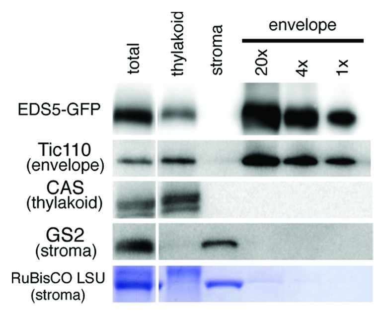
Figure 2. Immunoblot analysis of sub-chloroplast localization of EDS5-GFP. Chloroplasts isolated from 35S:EDS5-GFP plants were subfractionated into thylakoid membranes, stroma and envelope membranes. Fractions corresponding to 0.0065 mg chlorophyll were subjected to SDS-PAGE and immunoblotting analysis using anti-GFP (for EDS-GFP), anti-Tic110 (chloroplast envelope marker), anti-CAS (thylakoid membrane marker) and anti-GS2 (stroma marker) antibodies and anti-rabbit secondary antibodies. EDS5-GFP fusion proteins are specifically accumulated in the chloroplast envelope membranes.