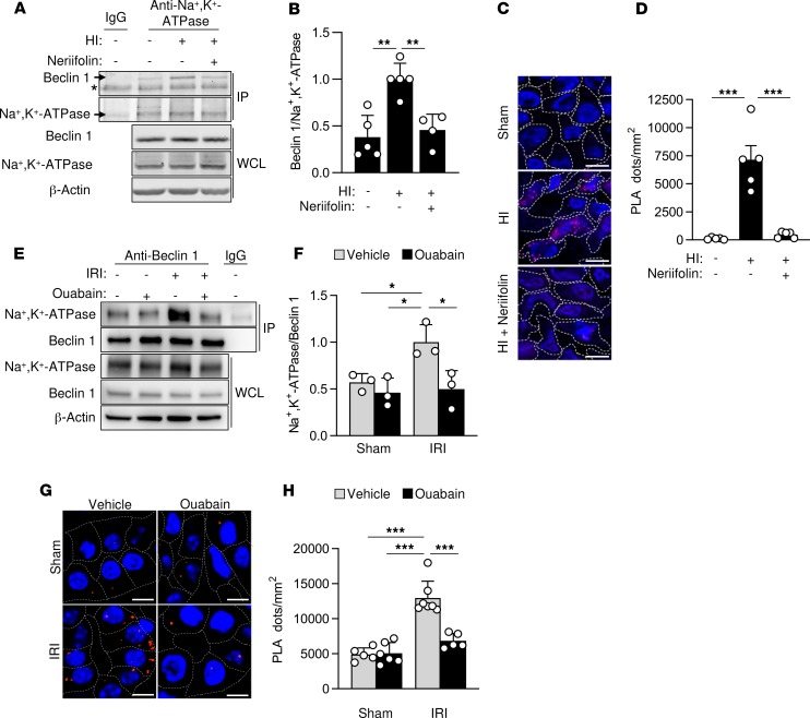
Figure 3. Beclin 1 and Na+,K+-ATPase interact in vivo during cerebral HI and renal IRI.
(A and B) Representative Western blots (A) and quantitation (B) of coimmunoprecipitation of Beclin 1 with the α subunit of Na+,K+-ATPase in the hippocampus of rat pups that were sacrificed 6 hours after receiving intraperitoneal vehicle or neriifolin (0.22 mg/kg) and subjected to sham operation or cerebral HI. In B, bars represent mean ± SD (n = 4–5 rats per group). (C and D) Representative images (C) and quantitation (D) of PLAs of Beclin 1 and the α subunit of Na+,K+-ATPase in the CA3 hippocampal region of rat pups that were sacrificed 6 hours after receiving intraperitoneal vehicle or neriifolin (0.22 mg/kg) and subjected to sham operation or cerebral HI. In D, bars represent mean ± SEM (n = 5 rats per group, 4 randomly selected fields analyzed per animal). (E and F) Representative Western blots (E) and quantitation (F) of coimmunoprecipitation of the α subunit of Na+,K+-ATPase with Beclin 1 in kidneys from mice that were subjected to sham operation or renal IRI after peritoneal administration with either vehicle or ouabain (0.25 mg/kg). In F, bars represent mean ± SD (n = 3 mice per group). (G and H) Representative images (G) and quantitation (H) of PLAs of Beclin 1 and the α subunit of Na+,K+-ATPase in kidneys from mice that were subjected to sham operation or renal IRI after peritoneal administration with either vehicle or ouabain (0.25 mg/kg). In H, bars represent mean ± SEM (n = 5–7 mice per group, 10 randomly selected fields analyzed per mouse). For A and E, the same tissue sample of the sham group (lane 1 of gel) was used as a control for IgG immunoprecipitation. Asterisk, nonspecific band. Scale bars: 10 μm (C), 50 μm (G). *P < 0.05, **P < 0.01, and ***P < 0.001; 1-way (B and D) or 2-way (F and H) ANOVA test.

