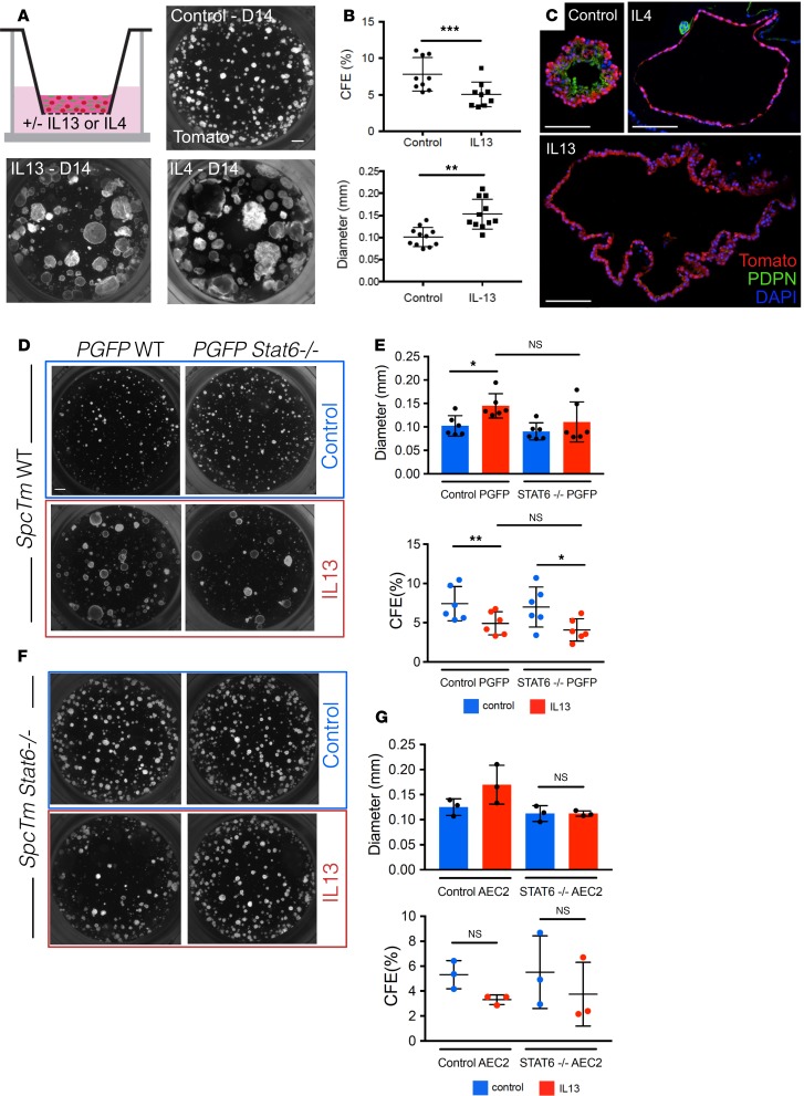
Figure 2. AEC2-derived organoids grown with IL-13 display altered morphology.
(A) 3D organoid cultures containing lineage-labeled AEC2s and PdgfraGFP+ stromal cells were grown with and without IL-13. Organoids grown with IL-13 appear larger and less dense. This morphology is observed with treatment with another Th2 cytokine, IL-4, as well. (B) When compared with control organoids, IL-13–treated organoids display lower colony-forming efficiency (CFE) and higher average diameter. Paired t tests; error bars indicate mean ± SD. (C) IL-13–treated spheres lack AEC1 staining (PDPN). Organoids treated with IL-4 display similar histological morphology. (D and F) Lineage-labeled AEC2s were isolated from WT SftpcTm mice (SpcTm WT) or from Stat6–/– SftpcTm mice (SpcTm Stat6–/–). PdgfraGFP+ stromal cells were isolated from WT PdgfraGFP mice or from Stat6–/– PdgfraGFP mice (PGFP Stat6–/–). These cells were plated in organoid culture in different combinations and grown to day 14. (D and E) Organoids derived from WT lineage-labeled AEC2s grow normally in control conditions, even if STAT6 signaling is abolished in the stroma. When IL-13 is added to WT organoids supported by either WT or Stat6–/– stromal cells, the typical IL-13 effect is observed. (F and G) Stat6–/– organoids grown with IL-13 do not display an increase in average sphere diameter, and the morphology appears grossly normal. These data suggest that the IL-13 effect is due to STAT6 signaling in the epithelium. One-way ANOVA; error bars indicate mean ± SD. Scale bars: 500 μm (A), 100 μm (C), and 500 μm (D). *P < 0.05; **P < 0.005.

