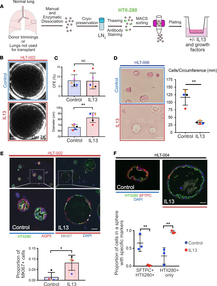
Figure 6. Human lung cell culture.
(A) Schematic for processing of normal human lung for culture of hAEC2s. (B) Representative example of human alveolospheres grown with and without IL-13. (C) CFE is similar with and without IL-13, but average sphere diameter is higher in IL-13–treated cultures. (D) Quantification of cell number per unit circumference. The cells in IL-13–treated spheres cover more surface area and appear elongated. Two-tailed t test; error bars represent mean ± SD. (E) Representative immunohistochemistry of AEC2s (HTII-280+) and AEC1s (AQP5+) in human alveolospheres grown with and without IL-13. Note the morphology of IL-13–treated spheres is different from controls and there is less staining for both AEC2 and AEC1 markers. There are more proliferating (MKI67+) cells in the IL-13–treated organoids. Two-tailed t test; error bars represent mean ± SD. (F) Representative spheres from a separate donor. Note larger diameter of IL-13–treated sphere and less expression of the AEC2 marker, SFTPC. One-way ANOVA; error bars represent mean ± SD. Dot colors correspond to separate donors. Donors are listed as HLT-xxx. Scale bars: 500 μm (B), 60 μm (D), 50 μm (E), 30 μm (F). *P < 0.05; **P < 0.005.

