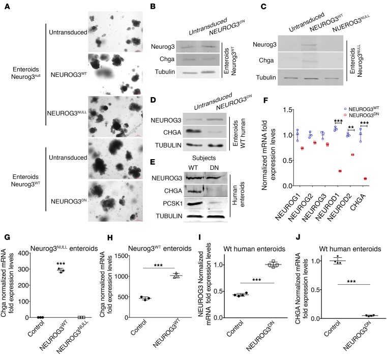
Figure 7. Assessment of endocrine cell induction in enteroids from Neurog3WT or Neurog3null mice or human intestines from normal and NEUROG3DN proband.
(A) Neurog3WT or Neurog3null enteroids transduced with either constitutively active NEUROG3WT- or NEUROG3NULL-expressing lentivirus and grown in culture for 7 days. (B and C) Western blot of enteroids cell extracts isolated from (B) Neurog3WT or (C) Neurog3null mice. Enteroids were untransduced or transduced with NEUROG3WT or NEUROG3NULL and assessed for proteins using anti-Neurog3, Chga and tubulin antibodies. (D) Western blot of enteroids cell extracts isolated from a normal human (WT) subject untransduced or transduced with NEUROG3DN and assessed 7 days later for proteins. (E) Human enteroids from WT and NEUROG3DN subjects were examined for specified proteins and (F) mRNA. (G and H) mRNA isolated from murine (G) Neurog3NULL and (H) Neurog3WT enteroids transduced with NEUROG3WT or NEUROG3NULL and WT enteroids transduced with NEUROG3WT and assessed 7 days later. (I and J) mRNA isolated from WT human enteroids and transduced with NEUROG3DN and assessed 7 days later. (F–J) *P < 0.05, **P < 0.01, ***P < 0.005, 1-way ANOVA and Dunnett’s multiple comparison. (G–J) *P < 0.05, **P < 0.01, ***P < 0.005, unpaired Student’s t test.

