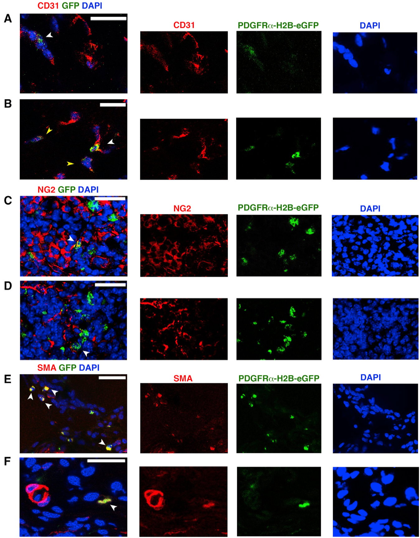
Figure 3. Differentiation Potential of PDGFRα+ Cells after Ischemic Insult.

(A–F) Seven days after HLI induction and adoptive transfer of undifferentiated GFP+PDGFRα+ cells, we tracked injected cells by nuclear GFP expression and co-staining with anti-CD31 (A and B), anti-NG2 (C and D) and Cy3-conjugated anti-αSMA antibody (E and F). White arrowheads indicate GFP+ cells; yellow arrowheads in (B) show decreased expression of GFP. Images representative of n = 3 independent experiments and were obtained with a confocal Leica SP5 DM as z stacks.
