
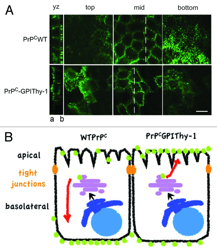
Figure 1. PrPC and PrPCGPIThy-1 expression in MDCK cells. (A) Confocal microscopy showing Z-stacks taken from top to bottom. As published before33PrPC (in green) is basolaterally (b) sorted in fully polarized MDCK cells whereas changing the SS-GPI of PrPC for the one of Thy-1 preferentially directs the PrPCGPIThy-1 to the apical (a) compartment. Scale bar is 10μm. Originally published in PLoS One. (B) Schematic representation of the differential sorting of wild type (WT) PrPC and PrPCGPIThy-1 shown in A.