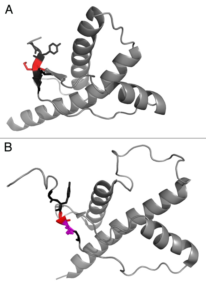
Figure 2. (A) Human PrP 90–230 structure from Zahn et al., 2000.61 PDB entry 1QM1. The sequence 127–131 is highlighted where residue 129 is colored red. (B) Chicken PrP 119–230 structure from Calzolai et al., 2005.62 PDB entry 1U3M. The sequence 127–131 is highlighted where residue 129 is colored red and residue 130 is colored in magenta. Sequence numbering according to human sequence.
