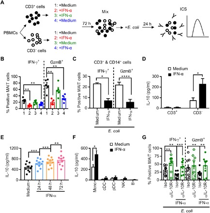
Fig. 5. IFN-α induces IL-10 production from monocytes and inhibits MAIT cell function.

(A) Schematic of the experiment. PBMCs from healthy donors were separated into CD3+ and CD3− populations, and each population was treated or not with IFN-α at 100 ng/ml for 72 hours. The CD3+ and CD3− cell populations from each donor were then mixed back together and stimulated by E. coli for 24 hours. (B) Cells were assessed for the production of IFN-γ and GzmB in MAIT cells. (C) CD3+ and CD14+ cells were purified from PBMCs and cultured together, followed by treatment with or without IFN-α for 72 hours. The cells were then stimulated with E. coli for 24 hours and assessed for the production of IFN-γ and GzmB in MAIT cells. (D) Levels of IL-10 in the supernatant of the CD3+ or CD3− population treated with or without IFN-α for 72 hours. (E) Level of IL-10 in the supernatant of PBMCs treated with IFN-α for 24 to 72 hours. (F) Levels of IL-10 in the supernatant of different subsets of PBMCs treated with or without IFN-α for 72 hours. (G) Levels of IFN-γ and GzmB expression in MAIT cells after a 72-hour treatment of PBMCs with IFN-α in the presence of an anti–IL-10R monoclonal antibody (αIL-10R; 10 μg/ml) or isotype control, followed by E. coli stimulation.
