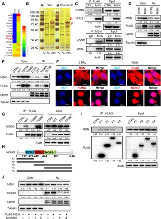
Fig. 4. Phosphorylated MSN enters nucleus to function for breast cancer progression with the assistance of a nuclear protein NONO.

(A) We used anti-FLAG M2 magnetic beads to carry out immunoprecipitation experiments on MDA-MB-231–CTRL and FLAG-MSN–overexpressing cell lysates and identified the protein samples by mass spectrometry. The protein components and relative abundances are displayed by a heatmap. (B) Immunoprecipitated samples by anti-FLAG M2 Magnetic Beads of CTRL or FLAG-MSN–overexpressing T47D and MDA-MB-231 cells were separated by SDS–polyacrylamide gel electrophoresis and silver staining. The red box shows differential bands, which appear in both cell lines. M, marker. (C) CTRL or FLAG-MSN–overexpressing MDA-MB-231 cells were immunoprecipitated (IP) by anti-FLAG M2 Magnetic Beads and then immunoblotted (top). Anti-MSN antibody was incubated with Dynabeads protein A and Dynabeads protein G and then to immunoprecipitated samples of MDA-MB-231 cells and immunoblotted (bottom). IgG, immunoglobulin G. (D) Cytoplasmic (Cyto) and nuclear (Nu) proteins were separated according to instruction. Western blot was conducted to determine the distribution of MSN in CTRL or FLAG-MSN–overexpressing MDA-MB-231 cells. Tubulin, internal reference for cytoplasmic proteins and lamin for nuclear proteins. (E) Western blot was carried out to determine the distribution of wild-type MSN and its mutants in MDA-MB-231 cells. (F) Immunofluorescence assay was carried out, in which endogenous MSN was determined with anti-MSN antibody in CTRL cells, exogenous MSN was determined with anti-FLAG antibody in FLAG-MSN–overexpressing cells, and endogenous NONO was determined with anti-NONO antibody. Images were captured by confocal laser microscopy. Scale bars, 10 μm. (G) CTRL or different-status MSN-overexpressing MDA-MB-231 samples were immunoprecipitated with anti-FLAG M2 Magnetic Beads and immunoblotted. DAPI, 4′,6-diamidino-2-phenylindole. (H) Schematic diagram of NONO in different truncated forms. (I) CTRL or FLAG-tagged different NONO fragment-overexpressing MDA-MB-231 cells were immunoprecipitated with anti-FLAG M2 Magnetic Beads and immunoblotted. (J) Cytoplasmic and nuclear proteins were separated and immunoblotted after MSN overexpression and NONO knockdown in MDA-MB-231.
